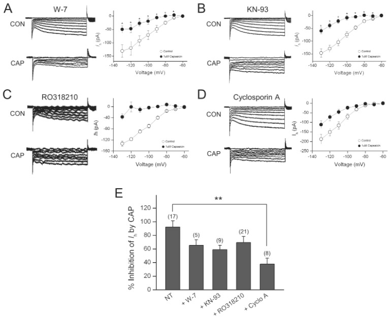
Fig. 5.
Cyclosporin A partially abolished the Ih inhibition by capsaicin. (A) Representative current responses of DRG neurons to 1-s voltage pulses under control condition, during application of 1 µM capsaicin (CAP) in the presence of W-7 in the pipette. Graph shows the current-voltage relationship of Ih under control and during capsaicin application in the presence of W-7. (B) Representative current responses of DRG neurons to 1-s voltage pulses under control condition, during application of 1 µM capsaicin in the presence of KN-93 in the pipette. Graph shows the current-voltage relationship of Ih under control and during capsaicin application in the presence of KN-93. (C) Representative current responses of DRG neurons to 1-s voltage pulses under control condition, during application of 1 µM capsaicin in the presence of thapsigargin in the pipette. Graph shows the current-voltage relationship of Ih under control and during capsaicin application in the presence of thapsigargin. (D) Representative current responses of DRG neurons to 1-s voltage pulses under control condition, during application of CAP in the presence of cyclosporine A in the pipette. Graph shows the current-voltage relationship of Ih under control and during capsaicin application in the presence of cyclosporine A. (E) Bar graph summarizes % inhibitions induced by capsaicin. Values at the top of each bar represent the number of experiments. Bars represent the mean±S.E.M. Asterisks indicate a significant difference from the first bar (**p<0.01).

