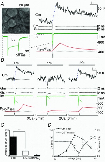
Figure 1. Ca2+ dependence of exocytosis in somata of MeV neurons.

A, left upper: photomicrograph of MeV neurons in a rat brain slice. Right: Cm jump evoked by a depolarizing pulse (from −60 mV to +10 mV, 100 ms). The Ca2+ currents (ICa) and {Ca2+}i with Ca2+ imaging (F340/F380) recorded simultaneously. The baseline Cm of the neuron was 53.02 pF (dotted line). The decay of the Cm response was fitted by a single exponential function with τ= 2.1 s (continuous line). B, pre-puffing 0 mm Ca2+ attenuated the Cm response (triggered by a depolarizing pulse to +10 mV, 100 ms). Both Ca2+ currents and {Ca2+}i decreased. C, statistical bar graph showing pulse stimulation-triggered Cm jumps as in B, that were abolished by 10 mm BAPTA in the pipette. D, Cm jump and Ca2+ current as a function of membrane potential. The Cm response was observable at membrane potentials positive to −30 mV, and peaked round 0 mV (n = 8). The Ca2+ current was recorded at different depolarizing potentials (100 ms, from −60 mV to 60 mV, +10 mV steps) in different cells (n = 8).
