
Figure 6. Pdk1 deletion rescues the synaptic plasticity deficit in Pten−/−mice.
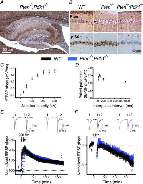
A, a representative image of immunohistochemistry for PTEN in hippocampal slices from Pten−/−;Pdk1−/− mice. As with Pten−/− mice, Pten is effectively deleted in most of the pyramidal neurons from the double-mutant mice, and their hippocampal architecture is normal. B, representative images of double immunostaining for PTEN and p-S6 in hippocampal slices of WT, Pten−/− and Pten−/−;Pdk1−/− littermates. The CA1 areas of the hippocampi are shown. C and D, basal synaptic transmission is not affected in Pten−/−;Pdk1−/− mice. Mean fEPSP slope as a function of stimulation intensity (C) or interpulse interval (D) measured at CA3–CA1 synapses in slices from WT or Pten−/−;Pdk1−/− mice are comparable. E and F, normal synaptic plasticity occurs in double-mutant mice. The mean fEPSP as a function of time before and after induction of LTP (E) and LTD (F) in slices from WT or Pten−/−;Pdk1−/− mice are similar. Insets show representative fEPSP traces before (1) and 180 min after (2) induction of synaptic plasticity.