
Figure 1. LiGluR evokes precisely timed and shaped Ca2+ rises in astrocytes.
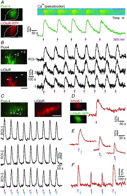
A, dual-colour TIRF image of a cultured cortical astrocyte transfected with LiGluR-RFP conjugated with the photoswitch MAG and loaded with the green-fluorescent Ca2+ indicator Fluo-4 (left). Dashed line shows the contour of the cell. The pseudocolour kymograph and green trace illustrate reproducible Ca2+ rises evoked by 385 nm light pulses (violet arrows, 0.3 mW mm-2, 50 ms). B, LiGluR photoactivation evoked repetitive and synchronized Ca2+ rises in astrocytic soma (ROI-1) and processes (ROI-2 and ROI-3). C, temporal shaping of astrocytic Ca2+ rises by switching LiGluR on and off with alternate 385 nm (violet arrows) and 488 nm (blue arrows, 39.1 mW mm-2, 200 ms) EPI light pulses. D–F, astrocytic Ca2+ elevations induced by LiGluR-GFP photoactivation and monitored with the red-fluorescent Ca2+ indicator Xrhod-1. The Ca2+ signals were shaped by switching off LiGluR with variable delay using 488 nm EPI light from the monochromator. Bars, 10 μm.