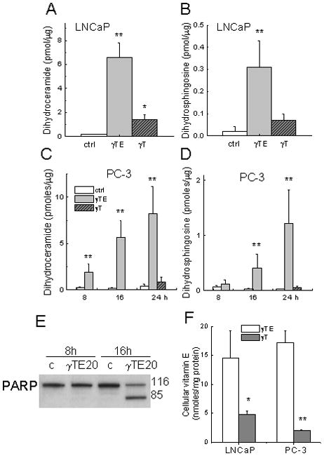
Figure 4.

Panels A and B - γTE (20 μM) or γT (50 μM) induced accumulation of dihydroceramide and dihydrosphingosine in LNCaP cells after 24-h incubation. Panels C and D - Effects on sphingolipids by γTE (20 μM) at indicated times or γT (50 μM, 24 h) in PC-3 cells. In Panels A–D, **P<0.01 and *P<0.05 indicate difference between γTE-treated and control cells. Panel E – Effects of γTE (20 μM) on PARP cleavage at indicated times in PC-3 cells. Panel F – Cells were incubated with γTE (10 μM) or γT (50 μM) in RPMI-1640 with 1% FBS for 6 h. Vitamin E forms extracted from collected cells were analyzed by HPLC with electrochemical detection. **P<0.01 and *P<0.05 indicate difference between γTE- and γT-treated cells.
