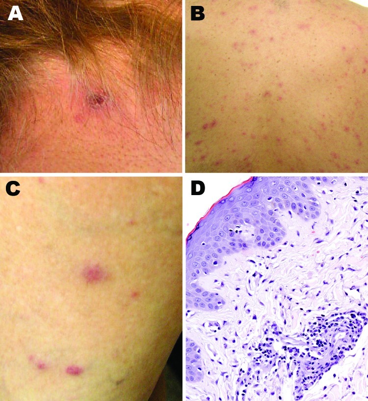
Figure 2.
Cutaneous lesions of patients with suspected and confirmed Rickettsia parkeri rickettsiosis in Argentina. A) Eschar at the nape of the neck at the site of recent tick bite. B, C) Papulovesicular rash involving the back and lower extremities. D) Histopathologic appearance of a papule biopsy specimen, showing perivascular mononuclear inflammatory cell infiltrates and edema of the adjacent superficial dermis and an intact epidermis (hematoxylin and eosin stain; original magnification ×100).

