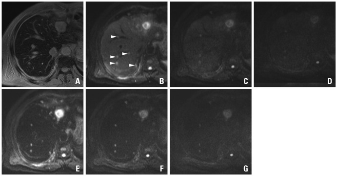
Fig. 5.
A 69-year-old man with colon cancer and numerous metastases throughout the liver including the largest one in the left hemiliver. On the SPIO-enhanced T2*-weighted image (A), metastatic lesions are not well distinguished from the background liver due to small lesion size and masking effect of the intrahepatic vasculature with high signal intensity. On the pre-contrast DWIs using b-factor (s/mm2) of 50 (B), at least 5 metastases (arrowheads) in a single level of right hemiliver are well delineated from the background liver due to 'black-blood' effect. The confidence is worsened on the images using higher b-factors of 400 (C) or 800 (D) with low signal intensity and marginal blurring of the lesions. On the SPIO-enhanced DWIs using b-factor (s/mm2) of 50 (E) or 400 (F), all five lesions are conspicuously seen, compared with poor marginal definition on the image using the b-factor of 800 (G). Compared with pre-contrast DWI, SPIO-enhanced DWI using corresponding b-factors shows consistently higher lesion-to-liver contrasts. SPIO, superparamagnetic iron oxide; DWI, diffusion-weighted MRI.

