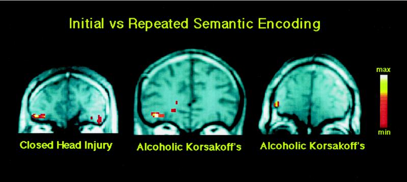
Figure 1.
Coronal view of prefrontal cortex in three amnesic patients with two different etiologies of amnesia. The slices, from left to right, are 39, 35, and 42 mm anterior to the anterior commissure. Individual activations are overlaid on T1-weighted anatomic sections. Each patient shows greater activation in the left inferior prefrontal cortex (corresponding to Brodmann area 47) for the initial relative to the repeated semantic processing of words. The individual activations correspond to the anterior extent of regions that show semantic activation in Fig. 3. The right side of the brain is depicted on the right side of Figs. 1–3.

