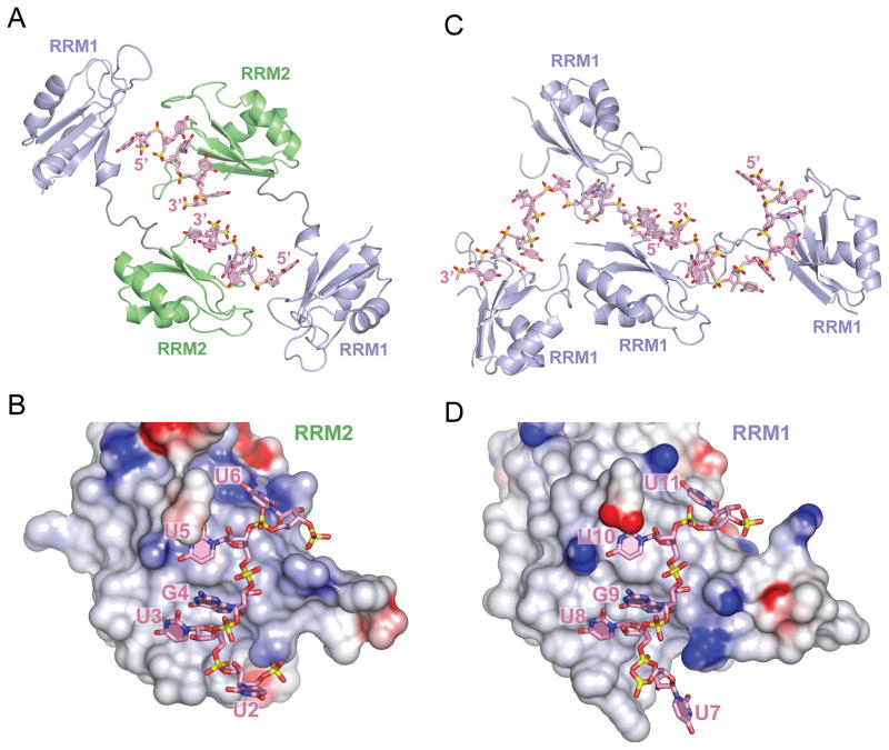
Figure 2. Overall Structures of CUGBP1 RRM1/2-RNA and RRM1-RNA Complexes.
(A) Ribbon and stick representation of two crystallographically related CUGBP1 RRM1/2 molecules bound to UGUU segments of two molecules of RNA GUUGUUUUGUUU RNA (sequence 1). The RRM2 domain (green) interacts with the U3-G4-U5-U6 segment, while RRM1 domain (blue) interacts with the U2 of the symmetry related RNA molecule. RNA strands are colored pink, with the backbone phosphorous atoms colored yellow. Nitrogen, oxygen and phosphate atoms are colored dark-blue, red and yellow in the RNA structure.
(B) An electrostatics surface view of RRM2 bound to U2-U3-G4-U5-U6 in the RRM1/2-RNA complex (RNA sequence 1) generated using the GRASP and PyMol programs. Basic and acidic regions of the protein appear in blue and red, with the intensity of the color being proportional to the local potential. The U3-G4-U5-U6 segment in pink (stick representation) contacts RRM2, while U2 is flipped out and directed away from RRM2.
(C) Ribbon and stick representation of four CUGBP1 RRM1 molecules bound to two molecules of UGUGUGUUGUGUG RNA (sequence 4) in the crystallographic asymmetric unit of the complex. Each RRM1 interacts with either U3-G4-U5-U6 or U8-G9-U10-U11 segment of the RNA. Protein and RNA are color coded as in panel A.
(D) An electrostatics surface view of RRM1 bound to U7-U8-G9-U10-U11 in the RRM1-RNA complex (RNA sequence 4). The U8-G9-U10-U11 segment in pink contacts RRM1, while U7 is flipped out and directed away from RRM1.
See also Figure S1.

