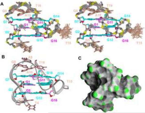
Figure 4. Solution Structure of the chl1 G-Quadruplex in K+-containing solution.
(A) Stereo view of 17 superpositioned refined structures. Guanosine bases in the G-tetrad core are colored cyan (anti) or magenta (syn). Bases in connecting loops are in biscuit color, with the backbone in grey and phosphorus and oxygen atoms in yellow and red, respectively. (B) Ribbon and (C) surface representations of a representative refined structure.

