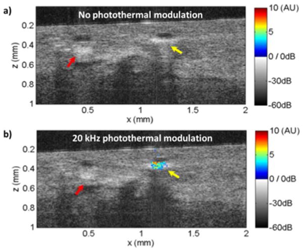
Fig. 4.

OCT images with (a) or without (b) photothermal modulation on freshly excised human breast tissue using injected nanorods as contrast agents. Photothermal OCT signal (in arbitrary units) with a SNR>5 is superimposed as pseudocolor on the gray scale OCT structural image. The yellow arrow indicates the injection site of the nanorods, while the red arrow indicates fibrostromal regions with strong backscattering.
