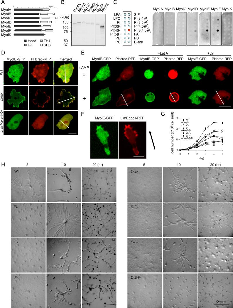
Figure 1. PIP3-binding class I myosins are required for normal cell growth and development.
(A) The domain structure of seven class I myosins in the Dictyostelium genome. (B) Immunoblotting of whole cell lysates prepared from Dictyostelium cells expressing myosin I-GFP fusions using anti-GFP antibodies. (C) Lipid dot blot assays show interactions of myosins ID, IE, and IF with PIP3. Images are representative of more than three independent experiments. (D) Localization of myosin IE-GFP and PHcrac-RFP in wild-type, pi3k-null, and pten-null cells (n ≧ 3, more than 10 cells analyzed in each experiment). Fluorescence intensity was quantified along the lines shown in Fig. S3. (E) Cells expressing myosin IE-GFP and PHcrac-RFP, a biomarker for PIP3, were observed before and after cAMP stimulation, in the presence or absence of latrunculin A or LY294002 (n ≧ 2, more than 25 cells analyzed in each experiment). (F) Cellular localization of myosin IE-GFP and LimEΔcoil-RFP, a biomarker for F-actin, in a cAMP gradient (n ≧ 6, more than 5 cells analyzed in each experiment). Arrow indicates the direction of cell migration. (G) Cell growth in wild-type and myosin I-null cells. Cells were counted daily. Values represent the mean ± SEM from more than four independent experiments. (H) Cells were plated on non-nutrient (DB) agar and examined for development. Images are representative of more than three independent experiments.

