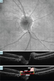
Fig. 2.
A horizontal B-scan OCT image crossing the center of the optic nerve head disc was extracted from a 3D square scan (, ). (a) The en face view of the optic nerve head. The artery, vein, cup, and optic disc are clearly visualized. (b) B-scan OCT image. The cup and disc are clearly visible in the horizontal cross-sectional OCT image (). (c) The B-scan OCT image () is marked to show the rim of the optic nerve and the cup-to-disc ratio (0.488).

