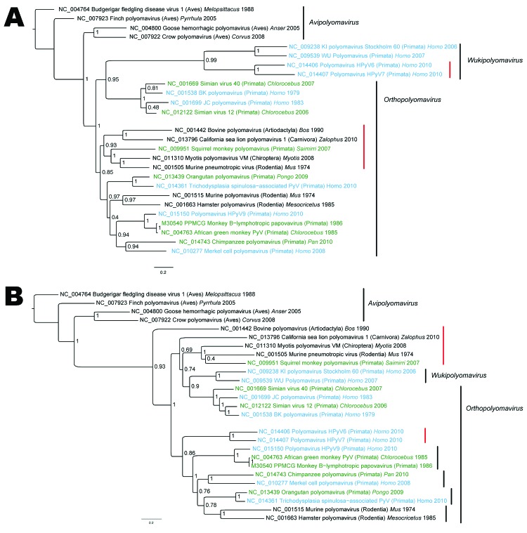
Figure 2.
A) Viral protein 1 (VP1) and B) large T antigen (LT) nucleotide-based phylogenetic reconstructions of polyomaviruises inferred by using a Bayesian method. Taxa annotations include reference number, name of the virus, host taxonomic order (in parentheses), host genus whenever available, and reported collection date. Human viruses are indicated in blue, and monkey viruses are indicated in green. Red vertical bars highlight groups for which VP1 and LT signals are incongruent. Posterior probabilities are indicated at each node. GenBank identification numbers are indicated directly on trees for each sequence. Scale bars indicate nucleotide substitutions per site.

