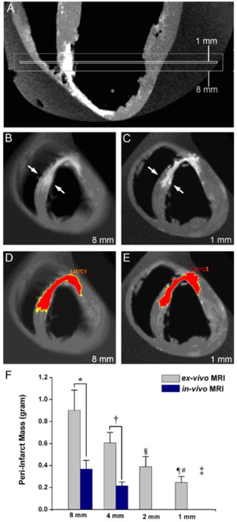
Figure 3. Peri-infarct zone assessment by ex-vivo MRI (n=5).
(A) Long-axis orientation showing the extent of 1 and 8mm slice thickness for short axis images. The presence of both viable and non-viable tissues in the 8 mm slice results in partial volume averaging. (B) For the 8mm slice thickness, the septal infarct appears transmural (arrows) and the PIZ is less defined with a wide range of gray values. (C) In the 1mm short axis slice at the same region it can be appreciated that infarct is not transmural and has well-delineated borders (D, E) The same myocardial slices with computer –generated mask depicting the core infarct (red) and PIZ (yellow). The PIZ is larger at 8mm. (F) Ex-vivo and in-vivo MRI showed different assessment of the PIZ at 8mm and 4 mm *p< 0.01 and †p= 0.01, respectively)
The PIZ mass in ex-vivo MRI acquisitions as a function of slice thickness showed marked differences (‡p=0.004) and decreased with thinner slice thickness; 2mm versus 8mm §p= 0.01, and 1mm vs 4 and 8mm, ¶p< 0.01 and #p< 0.001, respectively

