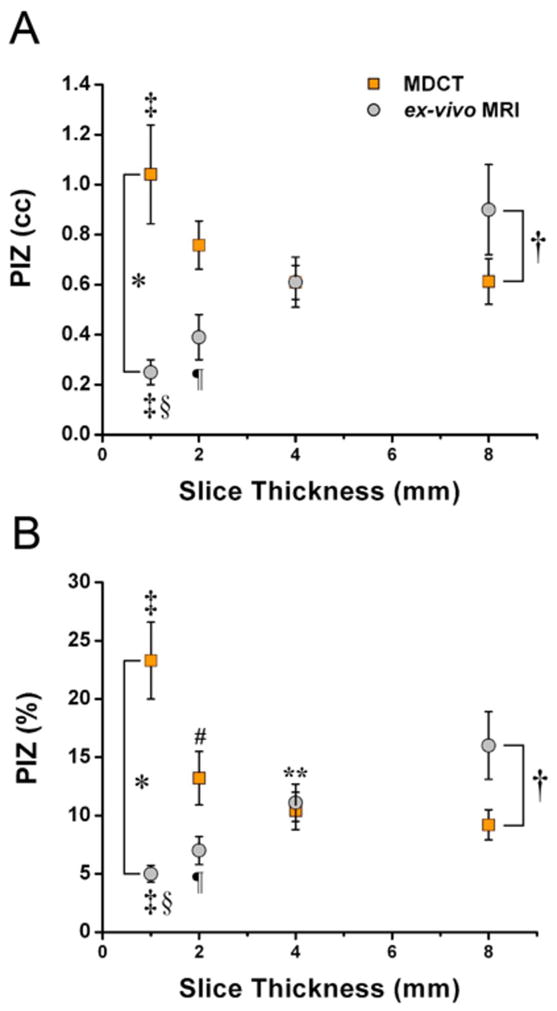
Figure 4. Comparison of PIZ assessment with ex-vivo de-MRI and de-MDCT.

The volume of the PIZ (A) and PIZ expressed as percentage of the total infarct size (B) decreases in a linear fashion with reduced slice thickness evaluated by ex-vivo MRI suggesting a pronounced partial volume effect, while the amount of PIZ volume is less affected by slice thickness in MDCT acquisitions until a slice thickness of 1mm is reached. This implies that MDCT assessment of the PIZ is less susceptible to partial volume effects, but affected by image noise at 1mm. There are marked differences in the PIZ volume and percentage assessment between MDCT and ex-vivo MRI assessment at 1 and 8mm *p<0.01 and †p<0.001, respectively. Differences between slice thicknesses: 1mm versus 4 and 8mm, ‡ p< 0.01 and §p< 0.001, respectively; 2mm versus 8mm, ¶p= 0.01 for ex-vivo MRI, and 8mm versus 2 mm 4 mm # p< 0.05 and **p< 0.05, respectively
