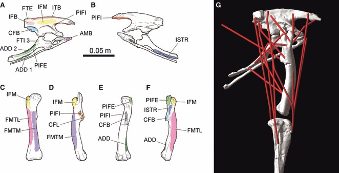
Fig. 2.

Myological reconstruction of the pelvis and hind limb of Lesothosaurus diagnosticus based on Maidment & Barrett (2011). (A,B) Pelvis in: (A) lateral; and (B) medial views. (C–F) Femur in: (C) cranial; (D) medial; (E) caudal; and (F) lateral views. (G) The 3D musculoskeletal model of Lesothosaurus in right lateral view. See Table 1 for muscle abbreviations. Scale bar equal to 0.05 m (a–f modified from Maidment & Barrett, 2011).
