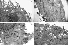
Figure 7.
Detection of cell ultrastructure with TEM after transfection. (a) SK-N-SH cells extended many cell filopodia toward a cluster of QDs (arrow) and simultaneously enveloped and phagocytized them (empty arrow). (b) The early endosomes (white arrow) are characterized as lipid droplets in the cytoplasm. The clusters of QDs were being phagocytized (arrowhead). (c) The early endosomes contained clusters of QDs at their edge (arrowhead). (d) The late endosomes resemble transparent droplets that include QDs and cell debris dispersed in the cytoplasm (white arrow). QD, quantum dots; TEM, transmission electron microscopy.

