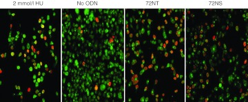
Figure 4.
Colocalization of PCNA and H2AX-r during gene editing. Confocal image of HCT116-19 cells synchronized with 2 µmol/l aphidicolin 24 hours beforeo the addition of 2 mmol/l Hydroxyurea, 72NT or 72NS, all at 8 µmol/l final concentration. After recovery, cells were stained for γ H2AX (red) and PCNA (green diffuse and punctate) to identify activated cells and viewed under confocal microscopy at 20× magnification.

