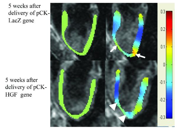
Figure 5.
Longitudinal strain in pixel-by-pixel obtained from phase contrast velocity encoded MR images in animals treated with pCK-LacZ and pCK-HGF gene. Longitudinal strain at diastole is shown in green (left images) and at systole in blue. Top block demonstrates longitudinal strain at diastole (left images) and systole (right images) at 5 weeks after infarction. At 5 weeks after infarction, a marked decrease in longitudinal strain was noted in the apex of both animals (arrow) compared with remote myocardium. An improvement in strain was noticed at 5 weeks after delivery of pCK-HGF (arrowhead, right image in the bottom block), but not pCK-LacZ (arrow) gene.

