
Figure 4.
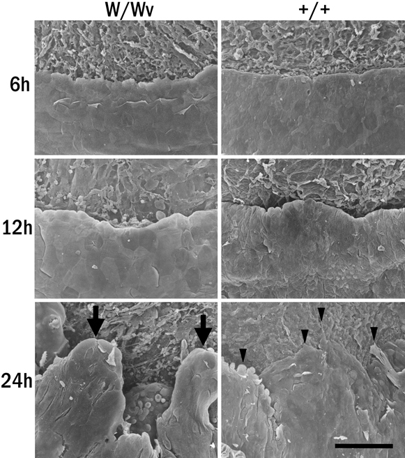
Scanning electron microscopy. The leading edges of the migrating cells of corneas from c-kit mutant and control WBB6F1+/+ mice, which were collected at 6, 12, and 24 h after epithelial wound healing, were examined by scanning electron microscopy. The leading edge of the c-kit mutant corneal epithelium at 24 h is elevated (arrows), whereas that of the WBB6F1+/+ makes flat lamellipodia (arrowheads) attachments on the cornea stroma. +/+=WBB6F1+/+ mice, W/Wv=c-kit mutant mice. Bar: 100 µm.