
Fig. 7.
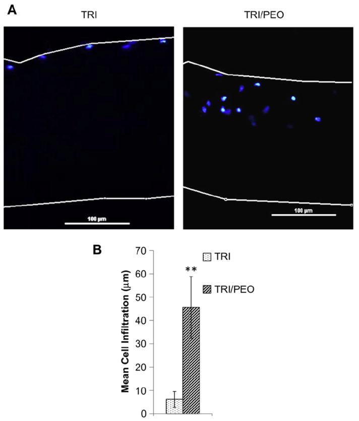
Electrospun scaffolds created with PEO fibers support cell infiltration of MSCs in vitro. After removal of PEO fibers from TRI/PEO scaffolds by washing, MSCs were seeded for one week. A) Scaffolds were sectioned and stained with DAPI to show cellular nuclei location. MSCs were able to infiltrate into the TRI/PEO scaffolds, but not TRI scaffolds, as seen by presence of nuclei within TRI/PEO scaffolds. B) Cell infiltration was quantified using a custom MatLab script. On average, MSCs seeded on TRI/PEO scaffolds migrated 45.59 μm into the scaffold, significantly greater than infiltration on TRI scaffolds (6.13 μm). An ** denotes p < .0001.