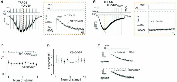
Figure 1. DrVSP-mediated inhibition (VMI) of TRPC6 currents.

A, current evoked by carbachol (CCh; 100 μm) recorded in the whole-cell mode from cells transfected with TRPC6 and DrVSP (left). Depolarization (+100 mV, 500 ms) was applied every 10 s (protocol displayed above). The area enclosed by the dashed box is enlarged in the right panel. Tail and outward currents are clipped to the frame here and throughout. B, data from cells transfected with TRPC6 and the enzyme-defective mutant DrVSPC302S. The conditions are the same as in A.C, averaged VMI elicited by repetitive depolarization in the presence of DrVSP (filled circles; n = 11) and DrVSPC302S (open circles; n = 5). D, averaged time constants of a single-exponential fit to the recovery from VMI. E, VMI of OAG (100 μm)- and RHC (100 μm)-induced TRPC6 currents (upper and lower panel, respectively). All VMI data are summarized in Fig. 2G and Table 1.
