
Figure 7. VMI in A7r5 aortic smooth muscle cells.
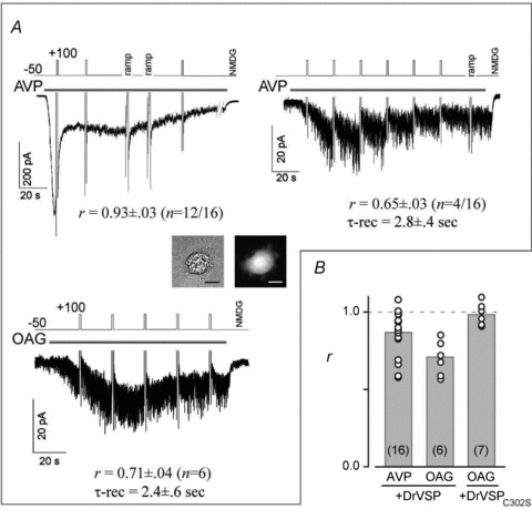
A, depolarization had almost no effect on TRPC6-like currents elicited in A7r5 cells by 50 nm AVP (left top, 12 of 16 cells). Co-transfection of the cells with DrVSP was confirmed by GFP expression (inset: bright-field (left) and GFP fluorescence (right)). A few cells exhibited clear VMI (right, 4 of 16 cells). OAG-induced currents also exhibited substantial VMI (left bottom). B, summary of VMI of AVP- and OAG-induced currents. VMI measured in each A7r5 cell (open circles); grey bars indicate the means of the VMI. Cells co-transfected with DrVSPC302S exhibited no VMI of OAG-induced currents.