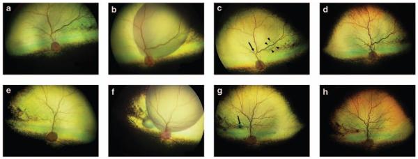
Figure 1.
Wide-angle fundus images (RetCam II, Clarity Medical Systems) of first and second subretinally injected eyes of a RPE65−/− dog (dog 6). The right eye (OD) was injected first and the left eye (OS) was injected 90 days later. (a) OD pre-injection, (b) OD immediately after subretinal injection, (c) OD 1-week post-injection. Note that in this eye, a small region of subretinal fluid remains (indicated by arrowheads in c), (d) OD 1-year post-injection, (e) OS pre-injection, (f) OS immediately after subretinal injection, (g) OS 1-week post-injection, (h) OS 1-year post-injection. The scars resulting at the injection site are indicated with arrows in (c, g).

