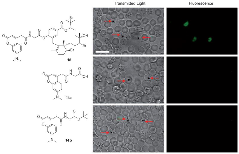
Figure 2.
Confocal microscopy images of live mixed-stage P. falciparum-infected erythrocytes incubated with either 10 μM bromophycolide A probe 15, 10 μM control tags 14a or 14b in complete medium. Hemozoin crystals were identified in the transmitted light image by dark crystals. Probe 15 localized only in P. falciparum-infected erythrocytes, as observed in the fluorescent image. No fluorescence was observed when P. falciparum-infected erythrocytes were incubated with either 14a or 14b. Red arrows indicate P. falciparum-infected erythrocytes. Echinocytes (altered red blood cells) are identified by the formation of small knoblike spicules on the membrane surface that cause the cell to transform from a biconcave disk into a seaurchin-like shape, an effect often observed with live cell imaging. Scale bar: 10 μm.

