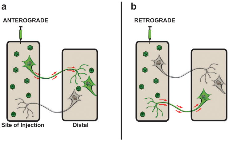
Figure 1. Anterograde and Retrograde transport after AAV infusion.
Diagram illustrates anterograde and retrograde transport of AAV vectors from the site of delivery to a second distally located brain region. (a) Axonal anterograde transport requires the transport of viral particles via an axon projecting from the site of vector injection to a distal area with subsequent transduction of cells located within the brain region where the axon ends. The presence of fibers only in the distal area is not classified as anterograde transportation of the AAV vector. (b) Retrograde transport of AAV vectors occurs when viral particles are taken up by axonal terminals in the injection site and are then transported back to the neuronal cell soma where they subsequently transduce the neuron.

