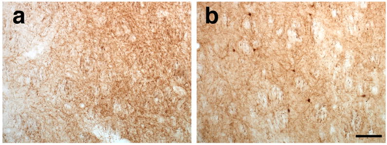
Figure 5. Thalamo-striatal projections corroborated the absence of anterograde transport of AAV6 vector 6 weeks after surgery.
Striatal analysis after thalamic AAV6 infusion reveals the absence of GFP+ cells (a) suggesting the lack of anterograde transport. In contrast, GFP-transduced cells were found in the striatum of animals that received AAV2 (b) demonstrating the anterograde transport of the vector. Scale bar 50 μm.

