
Figure 6. Retrograde axonal transport of AAV6 after striatal infusion.
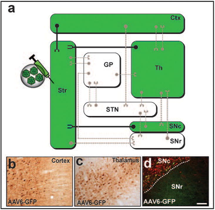
(a) Wiring diagram of the most representative connections in the basal ganglia organization. Transport of AAV6-GFP after striatal infusion is depicted in the diagram in green. Close examination of brain regions with known projections to the striatum demonstrated the presence of GFP+ cell bodies within the cortex (b), thalamus (c) and substantia nigra pars compacta (d, Green, GFP; Red, tyrosine hydroxylase), indicating a retrograde axonal transport of the vector through these projections. Note that green-colored regions in the diagram are representative of GFP+ cell bodies. Scale bar 500 μm.