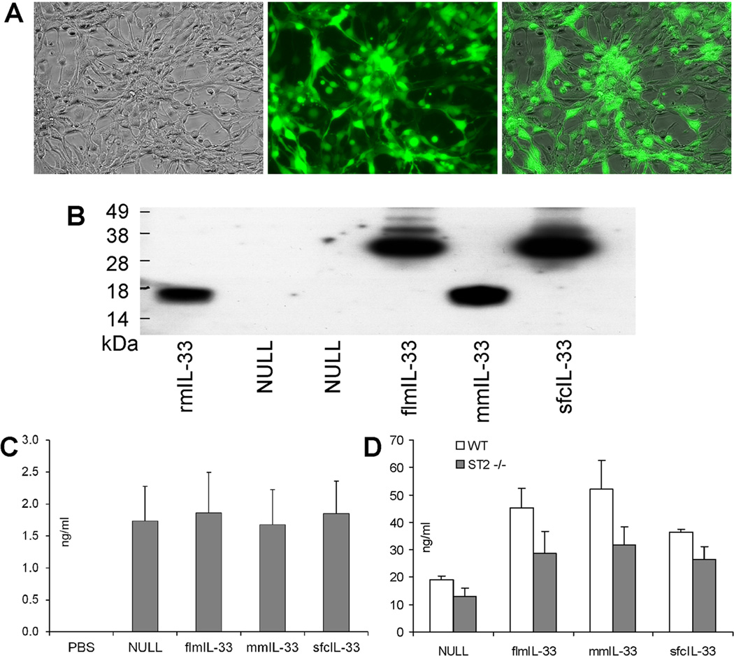
Figure 2.
Validation of infectivity and IL-33 gene delivery by recombinant AdV constructs. A. Recombinant adenoviruses infect cells causing green fluorescence due to GFP expression encoded in the viral backbone. Bright field microscopy (left), GFP fluorescence microscopy (middle), and image overlay (right) of AdV-flmIL-33-infected mouse epithelial TC-1 cells 48 h after infection (×20 objective). Similar results were obtained with all other constructs in these cells and also in A549 primary small airway epithelial cells and primary pulmonary mouse fibroblasts. No fluorescence was observed in cells without AdV infection. B. Western blotting with anti-mIL-33 antibody of fibroblast culture lysates infected with AdV encoding the indicated proteins. rmIL-33 is a commercial preparation of mature IL-33 (R&D Systems). Samples were normalized to total protein for loading. C. ELISA of whole lung homogenates for GFP, ng/ml ± SD, on day 14 after instillation of adenoviruses encoding the indicated proteins. D. ELISA of whole lung homogenates for mIL-33, ng/ml ± SD. WT mice (open bars) and ST2−/− mice (closed bars) were analyzed 14 days after infection with AdV encoding the indicated proteins, with 3–5 animals per group.

