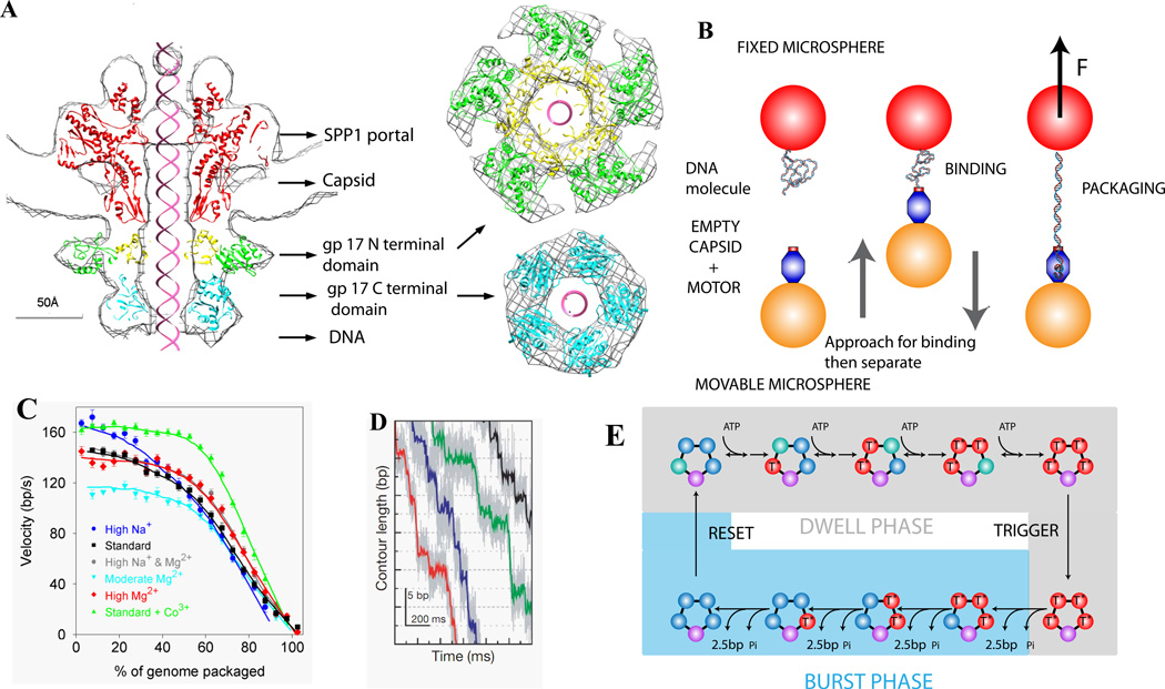
Figure 2.
(A) Structure of the Phage T4 DNA packaging motor [28]. (B) Measuring single phi29 viral DNA packaging using optical tweezers [34,35]. (C) Average motor velocity vs. % of genome packaged at 5 pN load. (D) High-resolution measurements of DNA translocation, showing phi29 packaging occurs in bursts of four ~2.5 bp steps [43]. (E) Schematic model of motor subunit coordination [43].

