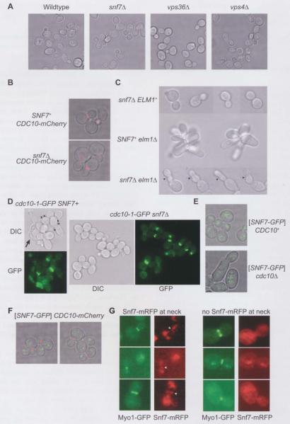
Figure 3.
ESCRT-III and septin localization patterns and effects on morphogenesis during normal and defective cell division.
Cells from exponentially-growing cultures were visualized by transmitted light microscopy of single focal planes (A–E) and either epifluorescence microscopy at regular intervals in the z direction combined with deconvolution of the resulting single focal plane images to eliminate out-of-plane fluorescence, followed by projection of each focal plane’s image onto a single image (B, E), or standard epifluorescence microscopy of single focal planes (D, G). (A) Strains were SEY6210 (wild-type), MWY24 (snf7Δ), MBY30 (vps36Δ), or MBY3 (vps4Δ) and were cultured at 30°C. (B) mCherry-tagged Cdc10, expressed from the endogenous CDCIO locus, was visualized with a rhodamine filter set in SNF7+ (JTY3992) or snf7Δ (YCS639) cells cultured at 30°C. (C) Cells of strain JTY4957 (snf7Δ ELM1), JTY4000 (SNF7+ elm1Δ), or JTY4973 (snf7Δ elm1Δ) pre-grown in YPD at room temperature were shifted to 37°C for 3 h. The arrowheads indicate constrictions along the lengths of snf7Δ elm1Δ buds. (D) Cells of strain JTY3986 (cdc10-1-GFP SNF7+) or JTY4000 (cdc1O-1-GFP snf7Δ), pre-grown in YPD at room temperature, were shifted to 30°C for 3 h. Cell morphology was visualized by differential interference contrast (DIC). Cdc10-1-GFP localization was visualized with an eGFP filter set (GFP). The arrowheads indicate cells with multiple attached buds. The arrow indicates an elongated, lysed cell. (E) Snf7-GFP was visualized with a TRITC filter set in cells of strain BY4741 (CDC10+) or YCS640 (cdc10Δ) cultured at 30°C and carrying plasmid pRS416-SNF7-GFP. (F) Snf7-GFP (green) and Cdc10-mCherry (red) were visualized in cells of strain JTY3992 cultured at 30°C using a TRITC or a rhodamine filter set, respectively. (G) Snf7-mRFP (red) and Myol-GFP (green) were visualized using a TRITC or an eGFP filter set, respectively, in diploid cells made by mating strain JTY3562 with strain JTY4510 and cultured at 30°C. Left column: cells with Snf7-mRFP at the bud neck (arrowheads); right column: cells in which no Snf7-mRFP was observed at the neck, imaged in the same fields as those in the left column.

