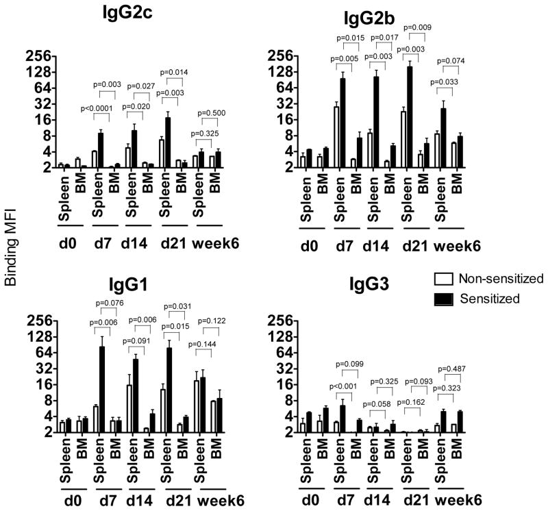
Figure 3. In vitro DSA secretion by spleen and bone marrow cells.
Spleen and BM cells were isolated from heart allograft recipients at 7, 14, 21 and at six weeks post transplant and cultured in vitro for 48 hours without re-stimulation. The presence of DSA was evaluated by testing the ability of the culture supernatants to bind donor thymocytes. Results are expressed as Mean Fluorescent Intensity (MFI). Spleen cells isolated from naïve B6 mice and cultured for 48 hours were used as a control. N = 4–5 mice per group at each time point.

