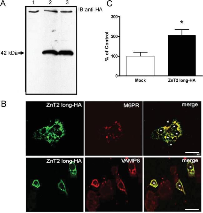
Figure 1. The ZnT2 long isoform is localized to the secretory compartment in mammary epithelial cells.
(A) Representative immunoblot of crude membrane proteins (100 μg protein/lane) from HC11 cells transfected with pcDNA3.1 (lane 1, mock-transfected control) or cells expressing the 42 kDa ZnT2 HA-fusion protein (lanes 2–3) detected with HA antibody. (B) Confocal micrographs of cells transfected with pcDNA3.1-longHA to express an HA-tagged 42 kDa ZnT2 fusion protein (ZnT2 long–HA) detected with HA antibody and visualized with Alexa Fluor® 488-conjugated anti-mouse IgG. Confocal micrographs illustrate that the long ZnT2 isoform (green) resides in distinct vesicular compartments and co-localizes (merge, yellow) with M6PR (red, endosome marker) and VAMP8 (red, exocytotic vesicle marker). Asterisks represent individual transfected cells (n = 3 and 6 cells expressing ZnT2 long-HA fusion protein). Bar, 20 μm. (C) Accumulation of Zn into vesicles was assessed using FluoZin-3 fluorescence in cells transfected to over-express the long ZnT2 isoform (ZnT2 long–HA) and compared with mock-transfected cells. Data represent mean percentage fluorescence relative to mock-transfected cells ±S.D. (n = 6–8 samples/group). *P < 0.05, a significant effect of over-expressing ZnT2 isoform on Zn vesicularization.

