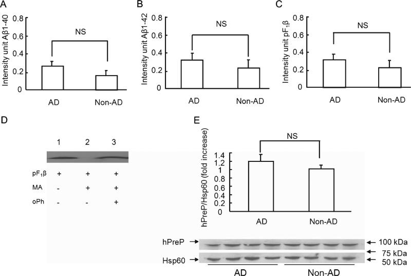
Fig. 2.
hPreP activity/expression in the cerebellum of AD and non-AD brains. Mitochondrial hPreP extracted from cerebellum for degrading biotin Aβ40/42 (A,B) and pF1β (C). Densitometry of Aβ or pF1β immunoreactive bands are shown by diagram bars. D) Effect of hPreP activity on degrading of F1β presequence (pF1β). pF1β was completely degraded by mitochondrial matrix hPreP protein (MA-hPreP, no F1β immunreactive band in lane 2 versus F1β immunoreactive band in lane 1 without MA-hPreP). In the presence of oPh, MA-PreP was not able to degrade pF1β (lane 3 versus lane 2 without oPh). E) Immunoblotting of cortical mitochondria from AD and non-AD cerebellum for human PreP. NS: no significant difference. Mitochondria were isolated from 12 AD brains and 8 non-AD brains. All experiments were performed in triplicates.

