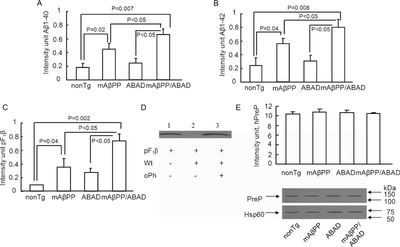
Fig. 3.
Alterations of PreP activity/expression in transgenic AD mice. Cortical mitochondrial PreP extracted from 5-month-old indicated Tg mice degraded the biotin Aβ40 (A), Aβ42 (B), and pF1β (C), respectively. Densitometry of Aβ40/42 or pF1β immunoreactive bands was performed using NIH image program. The upper panels indicate the representative immunoblots for Aβ (A, B) and pF1β (D). In the presence of oPh, mitochondrial PreP was not able to degrade pF1β peptide (lane 3 vs. lane 2). E) Immunoblotting of cortical mitochondrial protein from the indicated Tg mice for PreP. Representative immunoblots for PreP and Hsp60 are shown in the upper panel. Hsp60 (mitochondrial marker) was used as protein loading control and mitochondrial rich fractions. n = 4–7 mice per group. All experiments were performed in triplicates.

