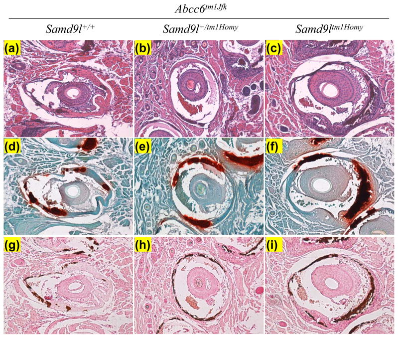
Figure 1.
Histopathologic examination of mineralization of the connective tissue sheath of vibrissae in Samd9ltm1Homy and Samd9l+/tm1Homy mice in comparison to their wild type counterpart (Samd9l+/+), all on Abcc6tm1JfK background. The stains used were hematoxylin-eosin (a–c), Alizarin Red (d–f), and von Kossa (g–i).

