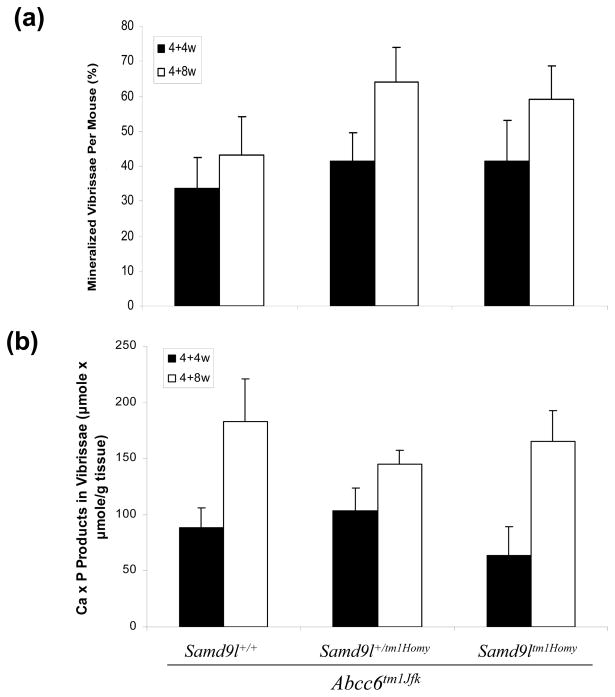
Figure 2.
Quantitation of mineralization of the connective tissue sheath of vibrissae in the group of mice shown in Fig. 1 by the count of mineralized vibrissae over total number examined (%) (a), and by chemical assay of calcium and phosphate in the skin biopsy specimens (b), at 4 weeks (4+4w) and 8 weeks (4+8w) of feeding with a diet accelerating the mineralization process in Abcc6tm1JfK mice. The values are mean + S.E., n = 6–13 mice per group.

