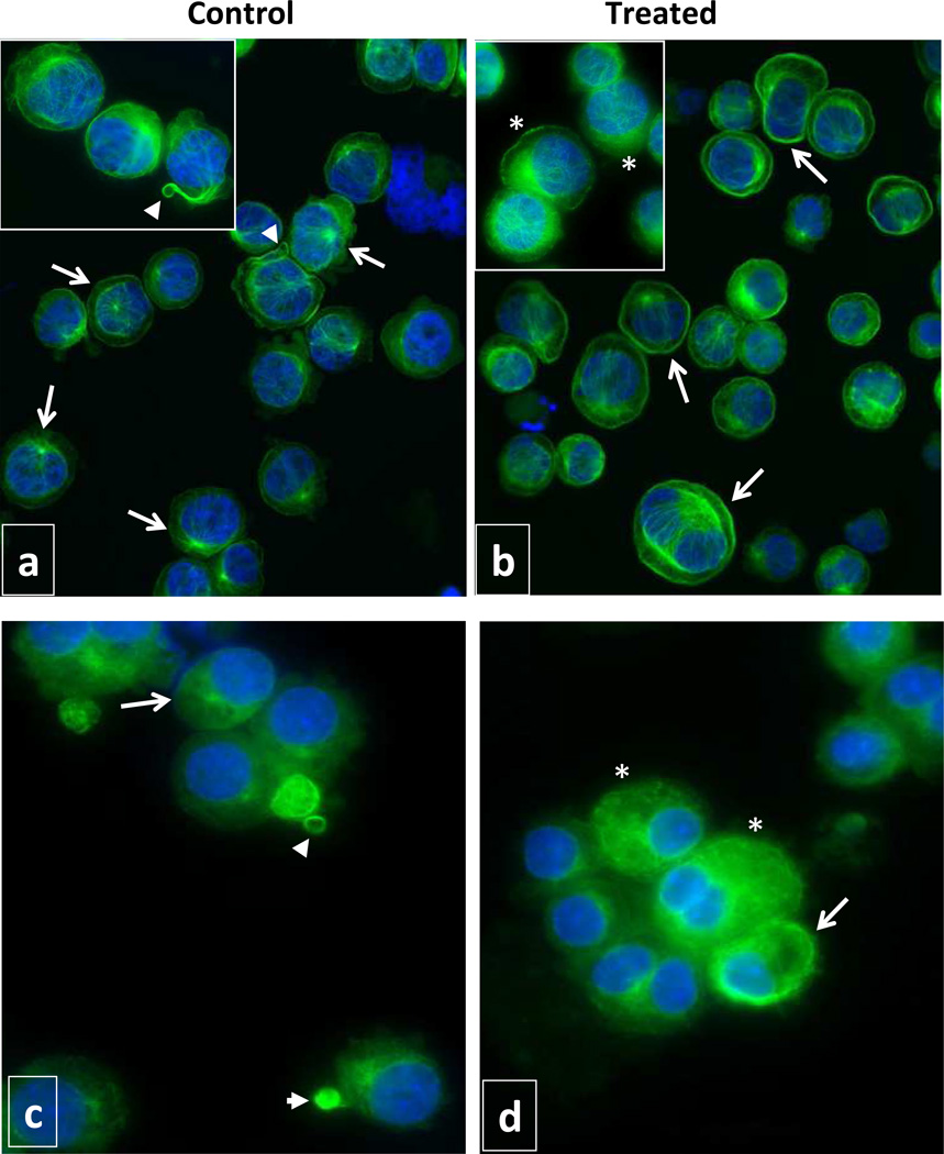
Figure 6. Evaluation of microtubule organization.
Immunofluorescence microscopy analysis of Meg-01 cells (a, b) and CD41+ primary MK (c, d) grown in the absence (a, c) and in the presence (b, d) of 2.5 nM LBH589 for 72 hours. The cells were labeled with anti-α-tubulin antibodies to visualize MT (green fluorescence) and with Hoechst 33342 to visualize the nuclei (blue fluorescence). Note, most of the cells in control cultures have normal network of MT radiating from the perinuclear MT organizing center (arrows in panels a and c) and some cells display MT structures resembling proplatelets and platelets (arrow heads in panels a and c). Most of the cells in LBH589-treated cultures have a marginal band of MT at the cell periphery or perinuclear (arrows in b and d), shorter filaments of MT or aggregates of polymerized tubulin (asterisks in b and d). Magnification 40X/0.60 dry objective in panels a and b; 100X/1.3 oil objective in inserts panels a and b; 63X/1.25 oil objective panels c and d.

