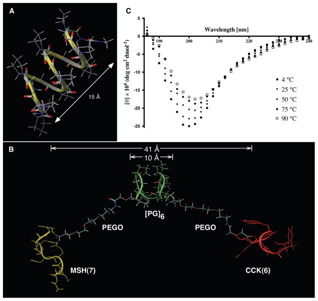
Figure 3.
Studies on linkers and heterobivalent ligand 12b. (A) Computationally generated structure of [PG]9 linker. (B) One of the conformations of htBVL 12b during MD simulation indicating the semirigid Pro-Gly backbone, the flexible PEGO ends, and β-turn features of MSH(7) and CCK(6) ligands with appropriate distances. (C) CD spectra of 100 μM of Ac-[PG]6-NH2 in water (pH 7) at different temperatures. The spectra reproduce a typical polyproline type II spectrum,27 albeit the positive band is not prominent.

