
Fig. 1.
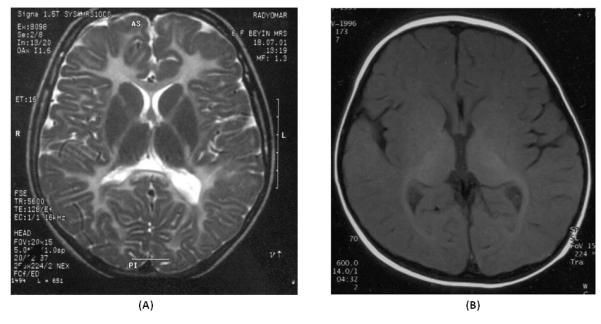
Brain MRIs of two patients analyzed in this study. a Axial T2-weighted image from patient F3.3 (with p.V182fs257X mutation in GJA12/GJC2) shows diffuse white matter hyperintensities with U fiber involvement. b Axial T1-weighted image from patient F4.3 (with PLP1 gene duplication) shows high signal intensity in the internal capsule, optic radiations like in a newborn indicating hypomyelination.