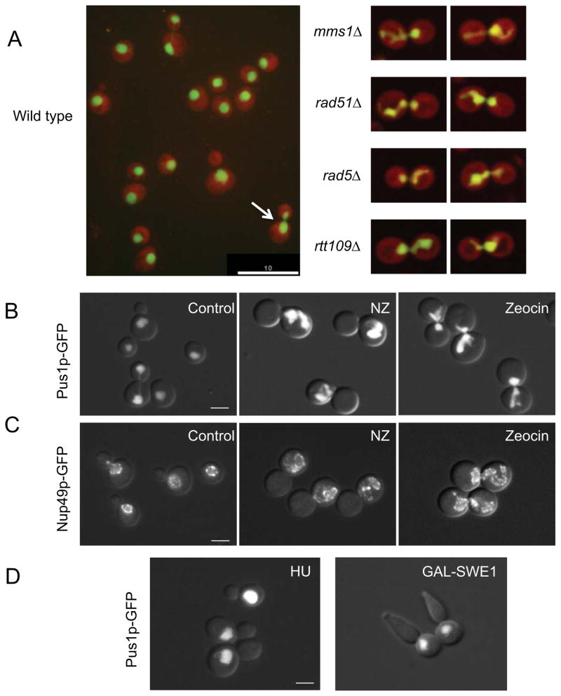
Figure 1. Mitotic delay leads to the formation of a nuclear extension.
(A) Images of strains from the automated microscopy screen. Cells are expressing a nuclear marker, Pus1p-GFP (green), and a cytoplasmic marker, TdTomato (red). Left: a field of wild type cells. The arrow points to a typical large budded cell at early stages of mitosis, with an elongated nucleus. Right: typical examples of cells of the indicated genetic backgrounds with nuclei exhibiting nuclear extensions. Scale bar =10μm. Additional images are in Supplemental Figure S1. (B and C) Wild type cells (KW913) expressing Pus1p-GFP (pane B) or Nup49p-GFP (pane C) were either untreated or treated with nocodazole (NZ) or zeocin for 3 hours. The nucleus in nocodazole treated cells does not traverse the bud neck due to the absence of microtubules. Scale bar =3μm. (D) Left panel: Wild type cells (KW926) were treated with hydroxyurea (HU) to induce an S phase arrest. Right panel: Cells over expressing SWE1 (MWY1152) and expressing Pus1p-GFP were arrested in G2 arrest by adding galactose. In both cases the arrest was achieved after three hours. Typical examples are shown. Scale bar =3μm. Quantification of the nuclear morphology of these strains and the control cells is presented in Table 1.

