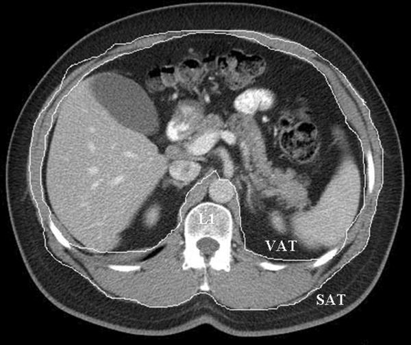
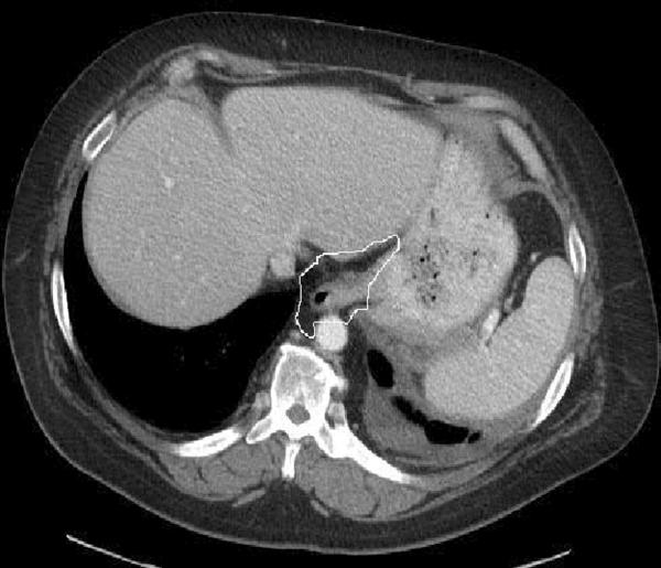
Figure 1. Representative cross-sectional images used for measurement of gastroesophageal junction (GEJ) and abdominal (visceral and subcutaneous fat).
a. Abdominal fat area: visceral and subcutaneous compartments, measured at level of the L1 vertebra.
b. GEJ fat area: measured without hiatal hernia.


