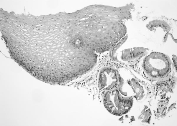
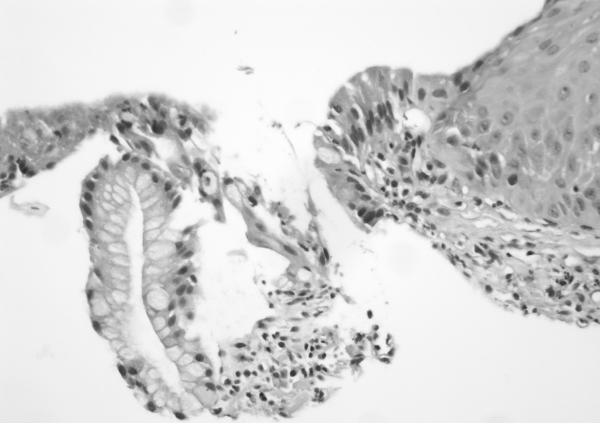
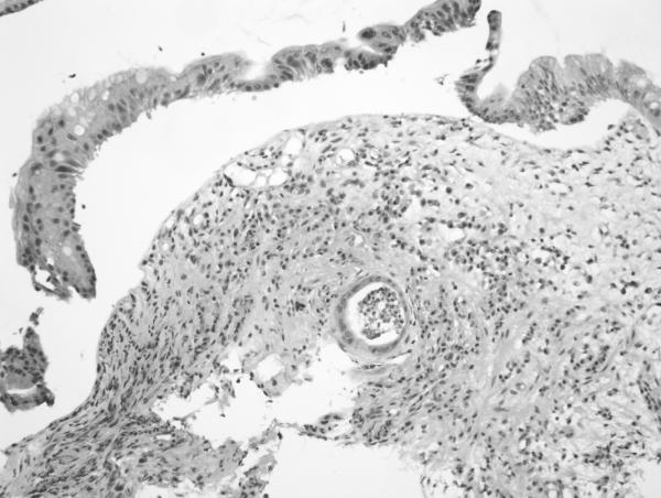
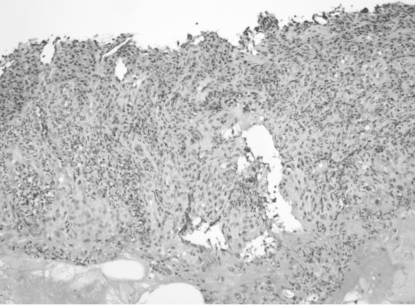
Figure 2. Grading of inflammation in BE biopsies from columnar mucosa. (on H&E sections).
a. Grade 0 (200×): Squamous mucosa (left) and Barrett mucosa (right) with no epithelial inflammation. The lamina propria also contains only a minimal amount of inflammatory cells.
b. Grade 1 (400×): High power view showing scattered epithelial infiltration by neutrophils within the Barrett mucosa (at left).
c. Grade 2 (200×): Greater inflammation than seen in Grade 1 cases. There is a crypt abscess in the lower half of the field as well as multiple foci of epithelial inflammation throughout the biopsy.
d. Grade 3 (200×): Ulceration of the mucosa with inflamed granulation tissue and abundant inflammation.


