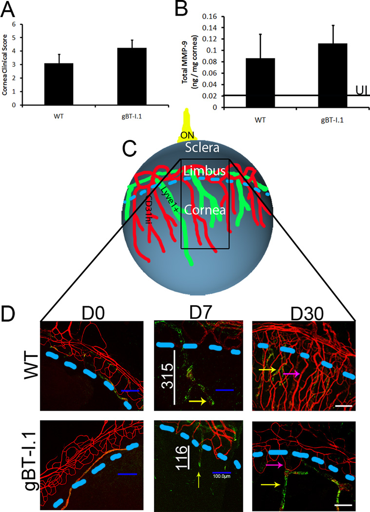
Figure 3. Ocular pathology in WT and gBT-I.1 mouse corneas.
WT and gBT-I.1 mice were infected with 1,000 PFU / eye HSV-1 McKrae. At 7 days pi, (A) clinical pathology was assessed and scored by a masked observer (clinical ophthalmologist) using a slit lamp exam and no significant differences were identified. (B) MMP-9 expression was evaluated by ELISA and were similar in gBT-I.1 and WT mice following infection. Data is represented as the mean ± SEM. Values represent 2 independent experiments of 2–3 mice per group. Black line, MMP-9 expression in uninfected controls. (C) An overview of the cornea following HSV infection. Blue line, edge of cornea (limbus region); black box, area imaged; ON, optic nerve. (D) Corneas were then assessed for lymphatic (green, Lyve1hi, CD31low) and blood vessel (red, CD31hi) growth into the cornea proper at 0, 7, and 30 days pi. Pink arrows, blood vessel growth; yellow arrows, lymphatic vessel growth; white lines, length of lymphatic vessel; blue dashed lines, edge of cornea (limbus region).

