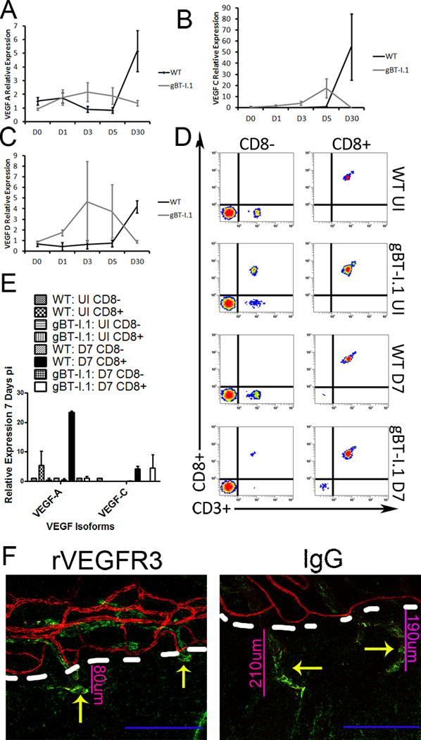
Figure 8. CD8+ T cells secrete VEGF-C to induce lymphatic growth into the cornea.
To evaluate the role of CD8+ T cells in lymphangiogenesis, mRNA transcript expression for VEGF-A (A), -C (B), and -D (C) was evaluated 0, 1, 3, 5, or 30 days pi in WT and gBT-I.1 corneas. To then confirm that CD8+ T cells were the major source of VEGF-C, CD8+ and CD8− cell populations were isolated from the draining MLN and spleen and evaluated for VEGF expression (E), and purity evaluated by flow cytometry (D). (E) To then substantiate the role of VEGF-C in lymphatic growth, chimeric VEGFR3 or control IgG was ocularly administered to gBT-I.1 mice following infection and lymphatic (green Lyve1hi) and blood vessel (red, CD31hi) growth evaluated by confocal microscopy. Dashed white lines, border of cornea proper with limbus.

