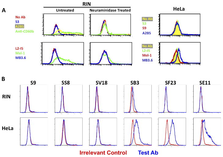
Figure 4. Binding of anti-CPSIII mAbs to cells expressing GD3/GT3 gangliosides on the cell surface.
Flow cytometry was used to study the binding of anti-GBS, anti-GD3, and anti-GT3 Abs to the surface of rat insulinoma cell line RIN and HeLa cells. Cell number is on the vertical axis, fluorescent intensity on the horizontal. (A) RIN cells were either treated with neuraminidase, or not, to demonstrate the sialic acid dependence of Ab binding; the HeLa cells were untreated. Binding of Abs was detected with FITC-conjugated isotype (murine IgM or IgG) specific Ab. IgM mAbs are at the top and IgG3 mAbs at the bottom in A. (B) Binding of anti-GBS mAbs to RIN (top) and HeLa (bottom) cells. Each graph compares binding of the test mAb (blue) to an irrelevant control mAb (red). FIGURE 4 SHOULD BE PUBLISHED IN COLOR.

