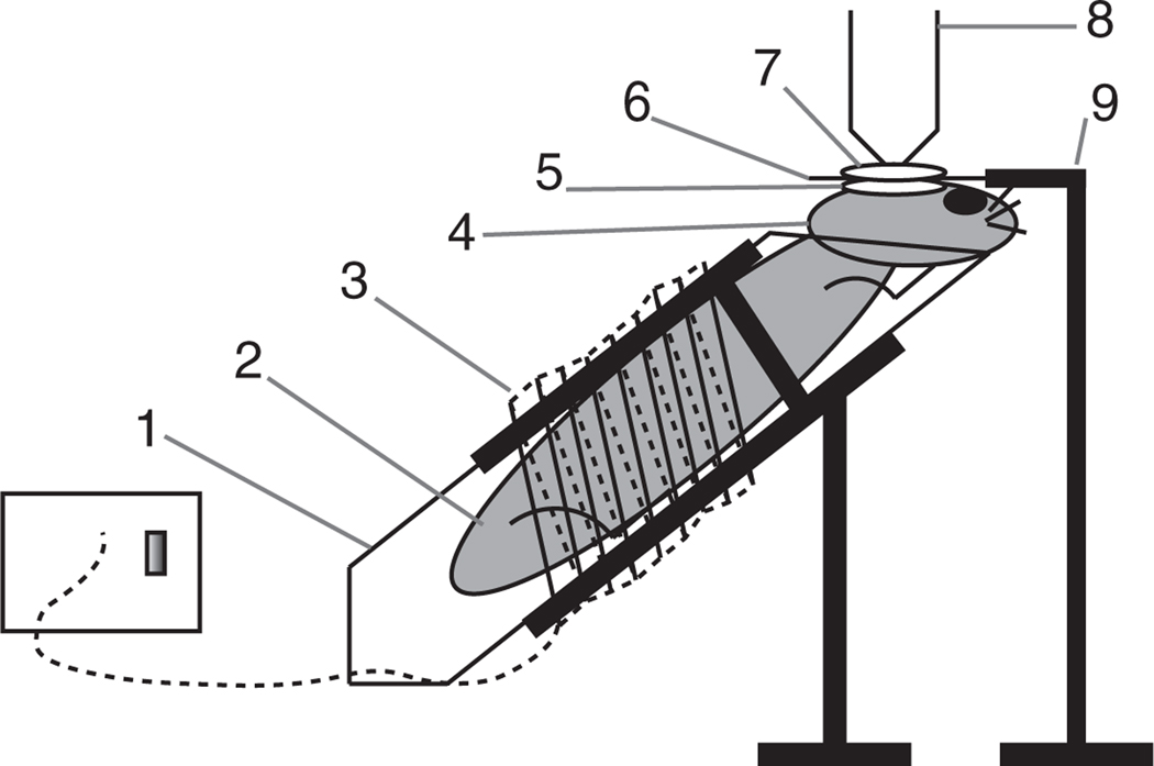
Figure 1.
Diagram representing a custom-made mouse imaging setup. (1) Mouse holder, obtained by modifying a 50-ml Falcon tube and placing it into a clamp/holder. (2) The mouse is positioned comfortably within the tube. (3) Heating module ensures that the mouse body temperature is kept at 37 °C. (4) The mouse head is positioned so that the imaged area is as horizontal as possible by resting the chin on the tube rim. (5) A drop of aqueous ointment or physiological saline solution is applied over the calvarium. (6) Cover slip. (7) A drop of water is placed over the cover slip. (8) Water immersion objective (alternatively, water dipping objectives can be used without the cover slip and can be placed directly onto the mouse calvarium). (9) The cover slip holder keeps the cover slip perfectly horizontal. Note: the drawing is not to scale. An isoflurane cone and scavenging system can be placed in proximity of the nose of the animal.

