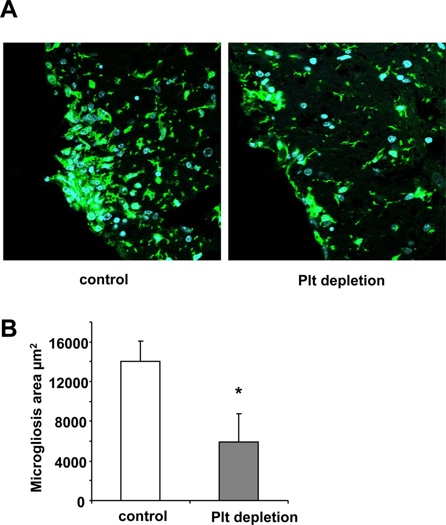
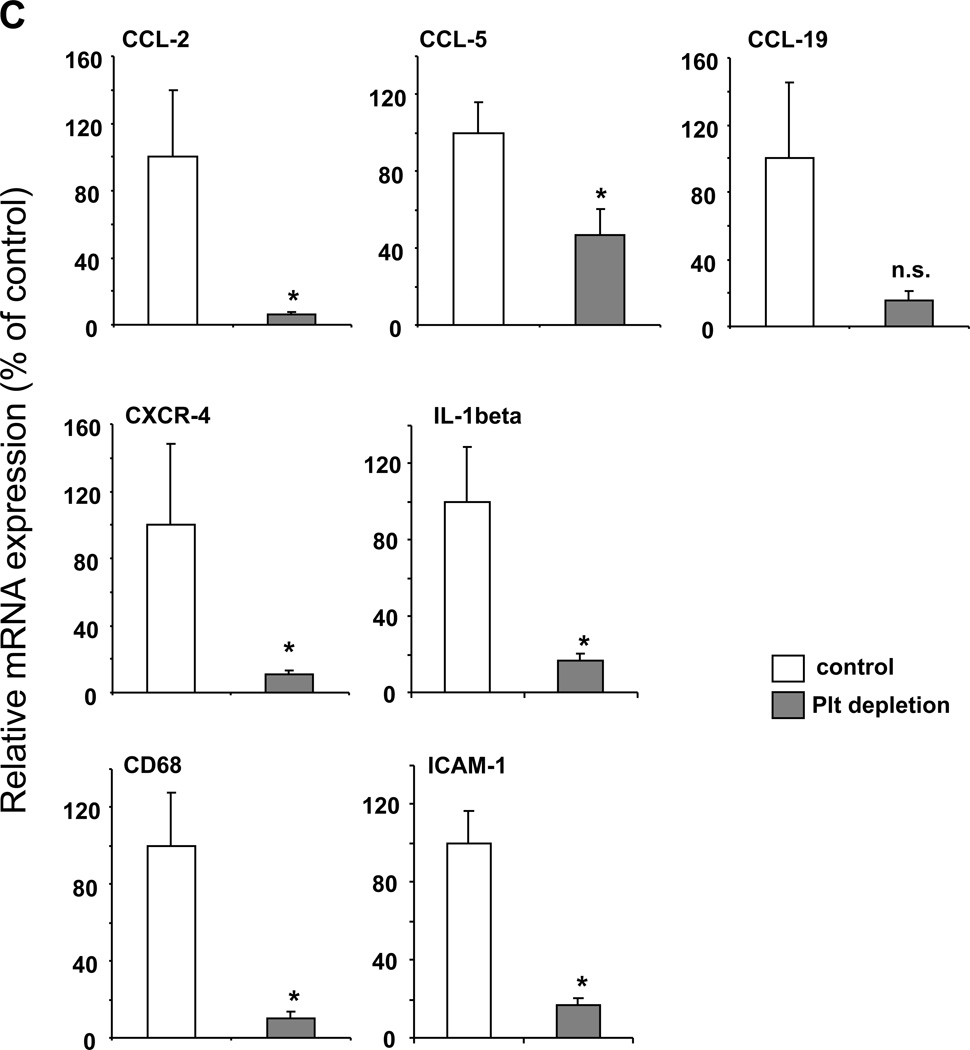
Figure 4. Platelet depletion reduces the inflammatory response during EAE.
To analyze the influence of platelet depletion on the inflammatory response in EAE, the disease was induced in WT mice and on days 12 and 16 post induction mice were treated with control serum (control) or with platelet-depleting serum. (A, B) On day 21, staining for microglia cells using the antibody to the marker Iba-1 was performed. The area of microgliosis was reduced in spinal cords upon platelet depletion. Representative images showing microgliosis areas are depicted. (B) The area of microgliosis was analyzed by morphometry. Six spinal cords per group and 3 non-consecutive sections per spinal cord were analyzed. Data are mean +/− SEM. * indicates p<0.05. (C) Spinal cords from control treated or platelet depleted mice were isolated on day 21 after induction of EAE. Real-time PCR analysis for the mRNA levels of the chemokines CCL-2, CCL-5 and CCL-19, the chemokine receptor CXCR-4, the cytokine IL-1β, the macrophage marker CD68 and the adhesion molecule ICAM-1 was performed. Data are mean +/− SEM (n = 5 mice per group) and are shown as percent of control. The mRNA expression of the respective molecule in spinal cords from control treated mice represents the 100 % control. * indicates p<0.05 as compared to control serum treated animals.


