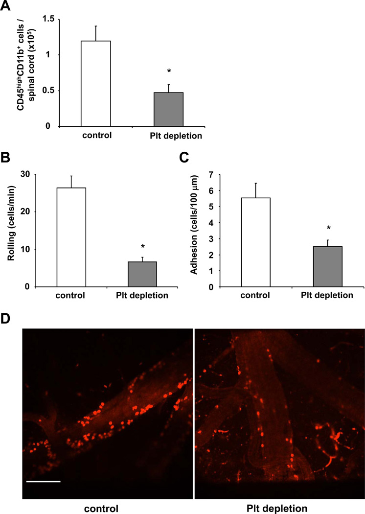
Figure 5. Role of platelets for leukocyte recruitment to the inflamed CNS in mice.
(A) EAE was induced in WT mice and on days 12 and 16 post induction mice were treated with control serum (control) or with platelet-depleting serum. Then, spinal cord tissue from WT mice treated with control serum (control) or with platelet depleting serum was collected after extensive systemic perfusion with saline on day 21 post immunization and leukocytes were isolated with percoll gradient centrifugation and analyzed by flow cytometry. The number of CD45highCD11b+ cells per spinal cord is shown. Data are mean +/− SEM (n=4–5 mice per group). * indicates p<0.05. (B, C, D) To test for the impact of platelets on leukocyte recruitment to the inflamed CNS, EAE was induced in WT mice and after 2 weeks mice, were treated without (control) or with platelet-depleting serum. Twenty-four h thereafter the number of labelled rolling (B) or firmly adherent (C) leukocytes to postcapillary venules was determined using spinning disc intravital microscopy. Data are mean +/− SEM and are shown as number of rolling leukocytes/minute or number of adherent leukocytes/100µm (n=3–4 mice per group). * indicates p<0.05 as compared to control. (D) Representative offline images of intravital microscopy experiments showing adherent leukocytes in control mice (left panel) or in mice after platelet depletion (right panel). Scale Bar: 100 µm.

